Running Head: Images in COPD: Endobronchial Valve
Funding Support: not applicable
Abbreviations: bronchoscopic lung volume reduction, BLVR; endobronchial valve, EBV; computed tomography, CT
Citation: Veselis CA, Steiner RM, Dass C. Bronchoscopic lung volume reduction using endobronchial valves: imaging appearance. Chronic Obstr Pulm Dis. 2020; 7(1): 60-63. doi: http://doi.org/10.15326/jcopdf.7.1.2019.0180
Introduction
Bronchoscopic lung volume reduction (BLVR) using endobronchial valves (EBV) provides an effective and safe alternative approach for reducing hyperinflation in patients with severe emphysema who are poor surgical candidates. Imaging plays a pivotal role in appropriate patient selection, target lobe identification and in the post-procedural management of adverse events.
History
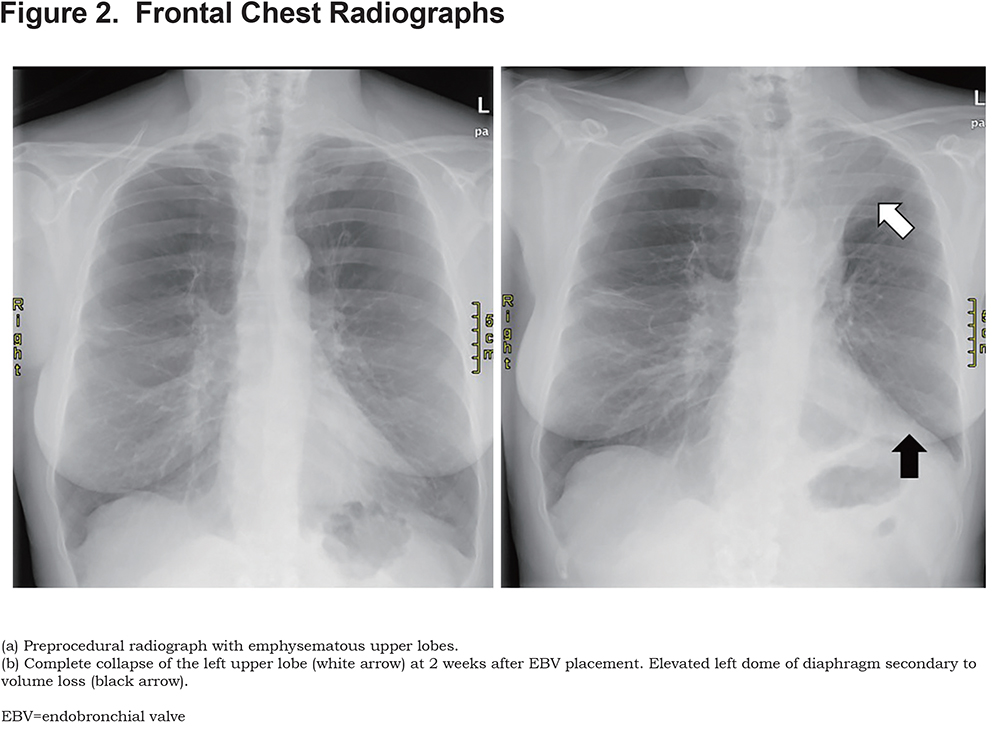
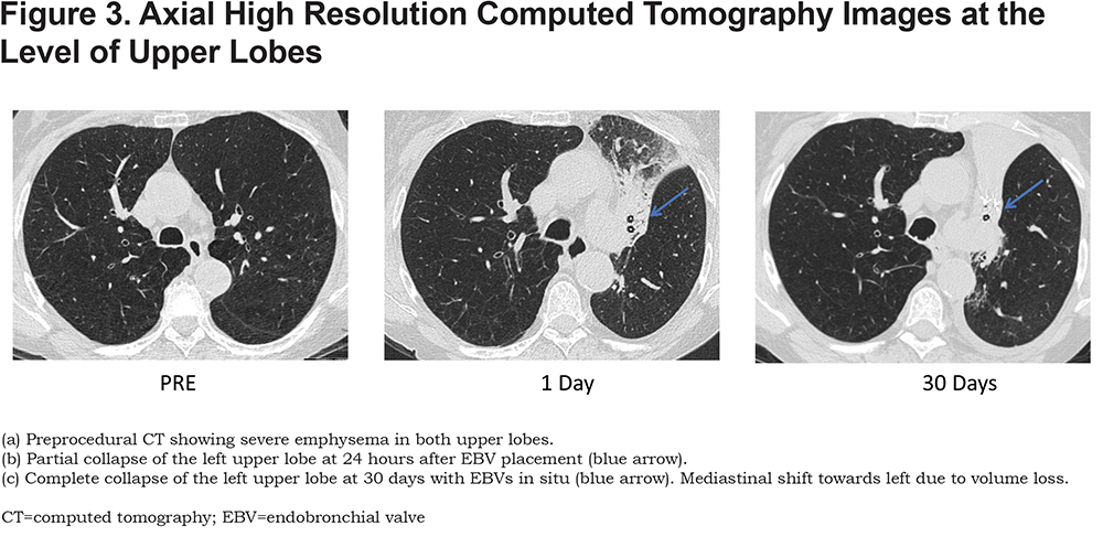
“A.B.” a 70-year-old female with a 50 pack-year smoking history and known severe emphysema presented for evaluation for BLVR. The patient had been previously managed medically, however, had persistent and worsening symptoms. The patient had been deemed a poor surgical candidate. After evaluation by the pulmonary team, A.B. was cleared for BLVR with one-way EBVs. A total of 5 EBVs (Spiration Valve System) were deployed in the left upper lobe segmental bronchus.
Imaging Findings
The following images were taken of patient AB. (Figures 1, 2 and 3)
Discussion
With the recent Food and Drug Administration approval of the Zephyr valve and the Spiration Valve System, BLVR using one-way EBVs is increasingly used in patients with advanced emphysema who remain symptomatic despite optimal medical management and are poor surgical candidates.1,2
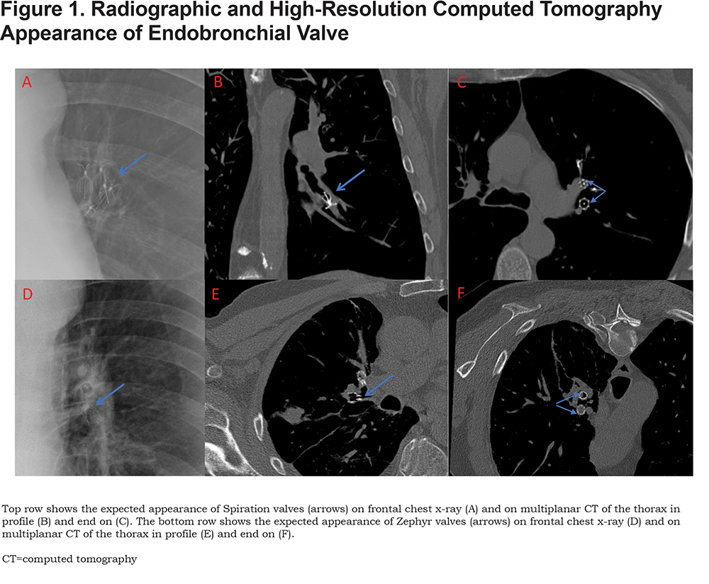
Given the higher incidence of pneumothorax after EBV placement, chest radiographs are required within 4 and 24 hours of EBV valve placement and daily thereafter until discharge. Knowledge of the types of EBV used, and the expected normal imaging findings following placement of EBV are essential to avoid misinterpretation of these imaging studies.
Significant volume reduction or atelectasis of the treated lobe may be observed within the first few days, although in some patients it may take up to a month. Criteria for premature reevaluation of these patients have been established and imaging plays an important role in their evaluation and management.3 An expert panel recommends a low dose computed tomography (CT) scan at around 30–45 days post-procedure to assess valve placement, particularly if there is no perceived improvement or breathing deterioration.3 The multidetector CT is evaluated by focusing on the valve count, location (migration/malpositioning) and the target lobe(s) total volume change (for either volume reduction or re-expansion after initial collapse).
Declaration of Interest
The authors have no conflicts of interest to declare at this time.