Running Head: Atopy and Serum IgE in Eosinophilic COPD
Funding Support: This study was funded by the National Institute of Environmental Health Sciences (R01ES031252).
Date of Acceptance: August 29, 2026 | Published Online Date: September 11, 2026
Abbreviations: BMI=body mass index; CI=confidence interval; COPD=chronic obstructive pulmonary disease; FEV1=forced expiratory volume in 1 second; FEV1 %pred=forced expiratory volume in 1 second percentage predicted; GOLD=Global initiative for chronic Obstructive Lung Disease; IgE=immunoglobulin E; IL=interleukin; IQR=interquartile range; OR=odds ratio; SD=standard deviation
Citation: Denoncourt CM, Elfeshawy MF, Ni W, et al. Allergic sensitization and serum immunoglobulin E in eosinophilic COPD. Chronic Obstr Pulm Dis. 2026; 13(5): 406-415. doi: http://doi.org/10.15326/jcopdf.2026.0770
Online Supplemental Material: Read Online Supplemental Material (682KB)
Note: The abstract of this paper was presented at the 2024 American Thoracic Society International Conference as a poster presentation with interim findings. The poster's abstract was published in 'Poster Abstracts' in the American Journal of Respiratory and Critical Care Medicine in 2024.
Introduction
Chronic obstructive pulmonary disease (COPD) is a heterogeneous condition characterized by persistent respiratory symptoms and airflow limitation, with a range of inflammatory phenotypes that influence clinical outcomes and treatment response. Among these, eosinophilic COPD has emerged as a clinically significant subtype, defined by elevated blood and sputum eosinophil counts and associated with type 2 airway inflammation.1 This phenotype is present in approximately one-third of individuals with COPD and has gained attention due to its increased risk of exacerbations and greater responsiveness to inhaled corticosteroids compared to noneosinophilic COPD.2 As research advances toward personalized approaches to COPD management, identifying and characterizing treatable traits, such as eosinophilic inflammation, has become increasingly important.
Despite growing recognition of eosinophilic COPD, important gaps remain in our understanding of its allergic features. The prevalence of allergic sensitization and elevated immunoglobulin E (IgE) is poorly characterized in patients with eosinophilic COPD despite the shared importance of eosinophilia and IgE elevation in type 2 immune responses.3 Exposure to aeroallergens in sensitized individuals is linked to poorer airway health (including greater symptoms, lower lung function, and more frequent exacerbations),4,5 and pharmacotherapies such as dupilumab that target allergic pathways have demonstrated the potential to reduce exacerbation rates in COPD. However, it remains unclear whether interventions to physically reduce these environmental exposures can improve outcomes, and determining the burden of allergic sensitization and IgE elevation in this subgroup, both with and without asthma and symptoms of allergic rhinitis, could have important implications for management of individuals with COPD and type 2 inflammation.
We conducted a cross-sectional analysis of baseline data from 174 former smokers with eosinophilic COPD enrolled in a randomized controlled trial of home air purification.6 We collected baseline measures of atopy and IgE as a supplement to the trial. Our aim was to determine the prevalence of allergic sensitization and elevated serum IgE in this population, and to examine the associations between self-reported allergen exposures, atopic conditions, and allergic sensitization. Better characterization of allergic sensitization within this population may inform future strategies for identifying, counseling, and treating patients with eosinophilic COPD in both research and clinical settings.
Methods
Study Design and Population
We included 174 former smokers with eosinophilic COPD who were recruited as part of a double-blind, randomized controlled trial of home air purification in Massachusetts.6 Participant recruitment included referrals from pulmonologists and primary care physicians, direct electronic medical record messaging, and outreach at clinical sites in Massachusetts. Skin prick allergen testing was conducted at the baseline study visit and represented the first formal allergen sensitization assessment for the majority of participants. Trial inclusion criteria were: (1) clinical diagnosis of COPD made by the patient’s treating pulmonologist or primary care physician, including Global initiative for chronic Obstructive Lung Disease (GOLD) Stage 2–4 airflow obstruction on clinical spirometry (forced expiratory volume in 1 second [FEV1] <80% of predicted and an FEV1/ forced vital capacity ratio <70%),7 (2) ≥10 pack-year smoking history and no longer smoking for at least 6 months, and (3) an absolute eosinophil count ≥150cells/μL in the past year. Disease status was adjudicated by review of medical records by the study’s clinical team. To reflect clinical populations with eosinophilic COPD, we did not exclude patients with a self-reported physician diagnosis of asthma. In the present study, only baseline data collected at the study entry visit, prior to trial randomization, are included. All participants provided informed consent to participate in the study. Institutional review was obtained through Beth Israel Deaconess Medical Center (2019P00129) and participating recruitment sites ceded review to this institution.
Data Collection
All study participants presented for a study entry visit at a clinical research center at Beth Israel Deaconess Medical Center in Boston, Massachusetts. All study participants completed a past medical history questionnaire in which they reported any history of asthma diagnosis by a physician, seasonal allergies (including history of allergic rhinitis or hay fever), and any hospitalizations in the past year due to COPD. Participants also reported cat or dog ownership and evidence of mice or cockroaches in the home, or moisture-related home conditions (water damage or dampness, or mildew/musty odor) via baseline questionnaire (Table S1 in the online supplement).
Lung function was obtained at baseline using a portable EasyOneTM Plus Diagnostic Spirometer (ndd Medizintechnik AG; Zurich, Switzerland).8 Allergic sensitization was determined via skin prick testing of 14 regional aeroallergens (Alternaria tenuis, Aspergillus fumigatus, dog epithelium, Cladosporium, cat hair, box elder, dust mite mix, cockroach mix, mouse epithelium, Penicillium, red birch, short ragweed, timothy grass, and white oak).9,10 A positive skin prick test was defined as an average wheal diameter ≥3mm larger than the negative control.10 Participants were not instructed to hold any of their home medications prior to the visit. Participants who were actively using portable air purifiers at home were excluded unless they agreed to discontinue their use for at least 3 months before enrollment. Central air conditioning and HVAC filtration systems were permitted. We also created categories of allergic sensitization: mammals (cat hair, dog epithelium, or mouse epithelium), fungi (Alternaria tenuis, Aspergillus fumigatus, Cladosporium, or Penicillium), grass/weeds (timothy grass, short ragweed), trees (box elder, red birch, white oak), and arthropods (cockroach mix and dust mite mix).
Total serum IgE was measured on the same day as skin prick testing. Elevated total IgE was defined as ≥100IU/mL, a clinically relevant threshold in the treatment of asthma that also has been associated with exacerbations and lung function decline in COPD.11
Statistical Analysis
We first performed descriptive statistics and determined the prevalence and overlap of allergic sensitization (to any of the 14 aeroallergens), asthma and seasonal allergies in this population with eosinophilic COPD. We then determined the prevalence and overlap of elevated IgE, asthma and seasonal allergies.
To determine if known aeroallergen exposure was associated with allergen-specific sensitivity, we then constructed logistic regression models to evaluate associations between self-reported exposure to cat, dog, mouse and cockroach at home and odds of positive skin reactivity to these specific allergens. We additionally examined associations between self-reported moisture-related home conditions—specifically, water damage or dampness in the home in the past 12 months and mildew or musty odor in the home—and fungal allergen sensitization (any of: Alternaria tenuis, Aspergillus fumigatus, Cladosporium, or Penicillium).
We tested if asthma diagnosis (COPD with asthma versus COPD without asthma), seasonal allergies, and serum IgE levels (≥100IU/mL versus <100IU/mL) were associated with higher odds of allergic sensitization through logistic regression models. Finally, to verify clinical significance in eosinophilic COPD, we tested if allergic sensitization (to any aeroallergens, specific allergens, and allergen categories), IgE levels, asthma, and seasonal allergies were associated with COPD hospitalization in the past 12 months using logistic regression models. We did not examine prospective COPD events due to the randomized controlled trial design. For all analyses, we adjusted for a prespecified set of potential sociodemographic confounders: age, sex, body mass index (BMI), and educational attainment (defined as completing at least some college). These specific variables were selected a priori due to their plausible associations with both environmental exposures and allergen sensitization patterns. We intentionally employed a parsimonious adjustment set to mitigate the risk of overfitting, given the limited number of outcome events in several of the allergen-specific models. Missing data were handled using complete-case analysis. Notably, covariate missingness was negligible: data were complete for age, sex and BMI, with education data missing for only a single participant.
To assess the robustness of our findings to further adjustment for lung function severity, we repeated primary analyses with the additional inclusion of baseline FEV1 (continuous, L) as a covariate.
Effect estimates were presented as odds ratios (ORs) with corresponding 95% confidence intervals (CIs). A 2-sided p-value<0.05 was considered statistically significant for all analyses. All analyses were conducted using R 4.4.2 (R Foundation; Vienna, Austria).
Results
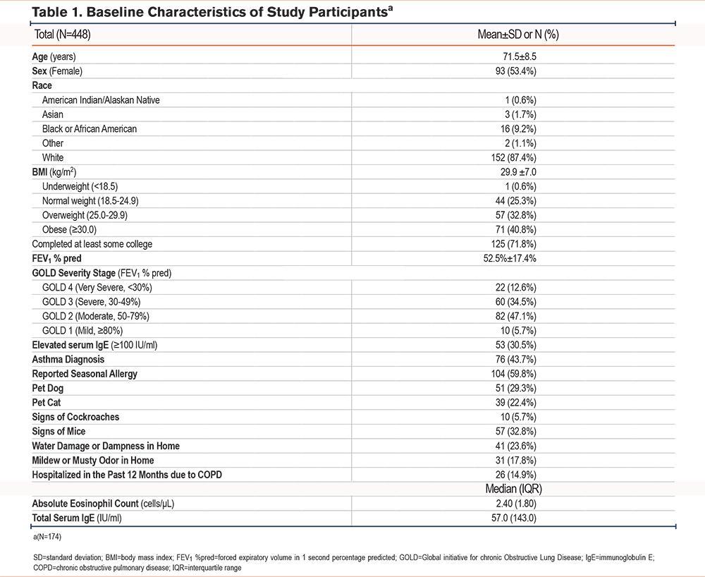
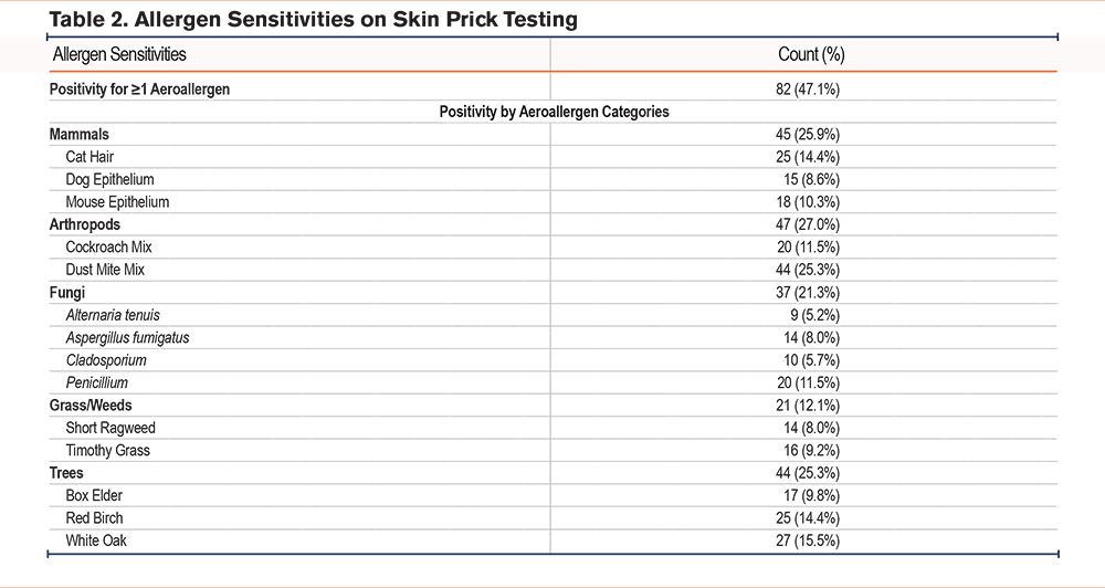
The study population is described in Table 1. There was a total of 174 participants with a mean ± standard deviation (SD) age of 71.5±8.5 years and an average FEV1 percentage predicted of 53.0% ± 17.3%. Overall, 14.9% reported a hospitalization in the past year due to COPD. Table 2 describes the prevalence of allergen-specific and overall skin prick sensitivity. Almost half of participants (47.1%) tested positive for at least one aeroallergen (allergic sensitization). Among 163 participants with IgE data, 32.5% had an “elevated” total IgE ≥100IU/mL. Overall, 55.7% (n=97) exhibited either allergic sensitization or elevated IgE. Seasonal allergies were reported by the majority of participants (59.8%), and 43.7% reported a prior diagnosis of asthma.
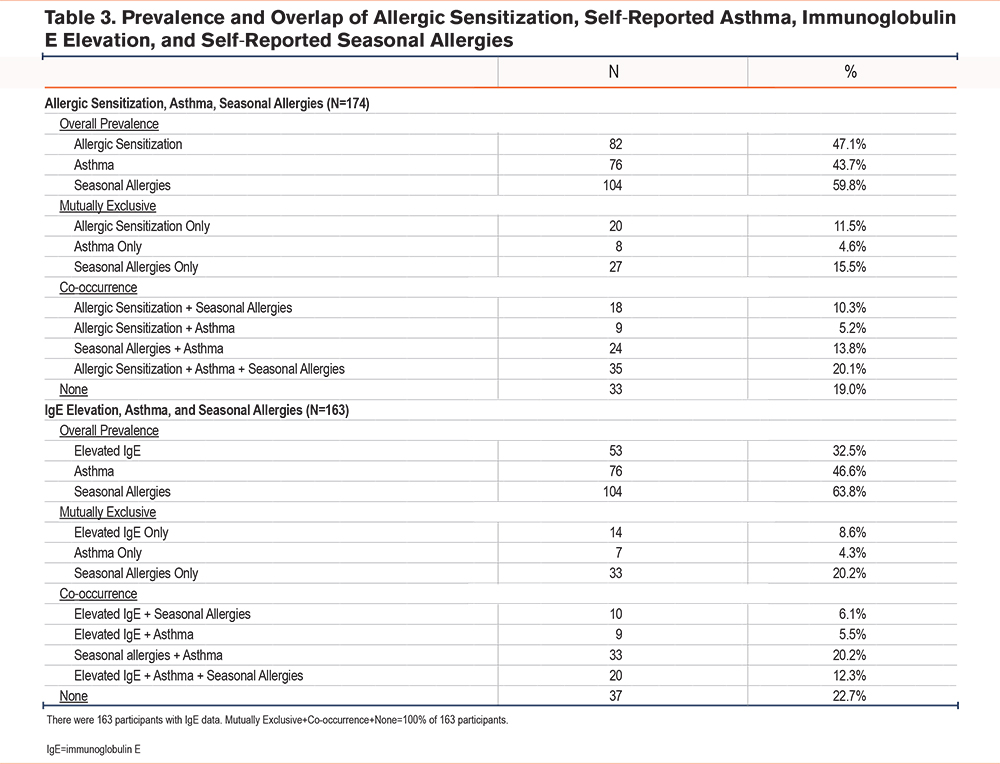
There was notable overlap of skin prick positivity with seasonal allergies, and asthma diagnosis in this population with eosinophilic COPD (Table 3). Even among those without an asthma diagnosis, allergic sensitization was common in this study population (38.8%). Only 19.0% did not have any skin prick positivity, asthma, or seasonal allergies. Similarly, there was overlap of elevated total IgE with seasonal allergies and asthma diagnosis (Table 3). Elevated IgE (≥100IU/mL) was common among those with asthma (42.0%), and also common among those without asthma (25.5%). Elevated IgE was detected in 31.3% of individuals with seasonal allergies and in 34.3% of those without seasonal allergies.
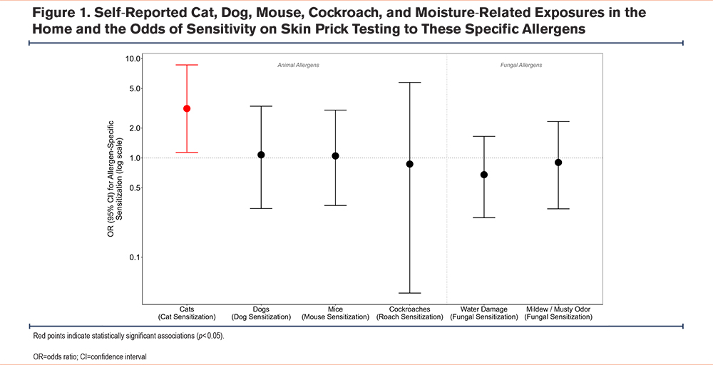
Of the 174 participants, 29.3% reported dog ownership, 22.4% reported cat ownership, 5.7% reported signs of cockroaches in their homes, and 32.8% reported signs of mice. Water damage or dampness was reported in 41 homes (23.6%), and mildew or a musty odor was reported in 31 homes (17.8%). Figure 1 shows associations between self-reported allergen exposures and the odds of allergic sensitization on skin prick testing. Cat ownership was associated with higher odds of allergic sensitization to cat hair allergen (OR=3.1 95% CI 1.1–8.6) in adjusted models. There was no association between self-reported dog ownership, signs of mice and signs of cockroaches, water damage or dampness, or mildew/musty odor and odds of specific allergic sensitization on skin prick testing.
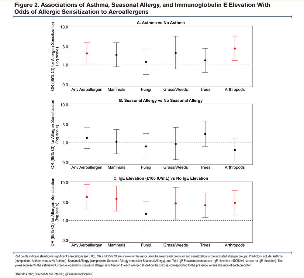
Asthma diagnosis (COPD with asthma) and IgE elevation were associated with allergic sensitization (Figure 2). Asthma diagnosis was a risk factor for nearly all specific skin prick sensitivities, including 2.0 (95% CI: 1.1–3.9) times higher odds of any aeroallergen sensitivity and a 2.7 times (95% CI: 1.3–5.8) higher odds of arthropods sensitivity. IgE levels were associated with allergic sensitization: patients with an IgE level ≥ 100IU/mL had 4.2 times higher odds (95% CI 2.0–9.0) of sensitization to any aeroallergen on skin testing compared to those with an IgE level <100. Associations between self-reported seasonal allergies and odds of allergic sensitization were less consistent (Figure 2). Associations with skin prick sensitivities to each of the 14 regional aeroallergens are detailed in Figure S1 in the online supplement.
Allergic sensitization, IgE elevation, and self-reported seasonal allergies were not associated with prior-year COPD hospitalization (Table S2 in the online supplement). Those with a history of asthma had higher odds of reporting COPD hospitalization in the past year (OR 1.6, 95% CI 0.7–4.2), although this association did not reach statistical significance. Allergic sensitization to animals, fungi, grass/weeds, or trees on skin prick testing showed no association with prior-year hospitalization.
Sensitivity analyses additionally adjusting for baseline FEV1yielded results consistent with our primary findings (Table S3, Figures S2 and S3 in the online supplement).
Discussion
In this study of older adults with eosinophilic COPD, we found a high prevalence of allergic sensitization and elevated IgE. The prevalence of allergic sensitization and IgE elevation in COPD has been described12 between 15%-40%, and we find nearly 56% of people with eosinophilic COPD in our cohort had these atopic markers. Exposure to cats in the home was associated with allergic sensitization to cat hair, while self-reported exposure to dogs, cockroaches, and mice was not associated with specific sensitization. As expected, our study found that asthma history is associated with atopy, however, we also found a high prevalence of allergic sensitization in those with eosinophilic COPD in the absence of an asthma diagnosis.
Atopy in COPD
Our findings align with several population-based studies that have demonstrated the presence of allergic sensitization and elevated IgE in subsets of individuals with COPD. In the COPDGene cohort, over 30% of participants with COPD had evidence of allergic sensitization based on serum-specific IgE, with sensitization associated with increased exacerbation frequency and symptom burden.13 Similarly, data from the SubPopulation and InteRmediate Outcome Measures In COPD Study (SPIROMICS) indicated that allergic sensitization was more common in younger individuals with COPD and was associated with worse respiratory health.13 The relatively advanced age of our cohort (mean age 71.5 years) should also be considered when interpreting these findings. Aging is associated with immunosenescence, including age-related alterations in humoral immunity and IgE responses, which may reduce the magnitude of allergic sensitization despite ongoing exposure. Consequently, the prevalence of allergic sensitization observed in this older cohort may underestimate the burden of atopy in younger individuals with eosinophilic COPD.
We did not find that allergic sensitization or IgE elevation was associated with hospitalization for COPD in the previous year. Other studies have found atopic status in COPD patients is associated with an increased incidence of respiratory symptoms such as cough and phlegm.14 These findings suggest that atopy may influence the frequency and severity of respiratory symptoms in COPD patients, potentially impacting their overall disease progression and response to treatment. Prospective studies are needed to evaluate if environmental allergies predict respiratory symptoms in eosinophilic COPD, in which case more screening, treatment, and environmental mitigation may be warranted.
Asthma Overlap
Asthma was reported in a subset of participants within our cohort and was notably associated with higher total serum IgE levels and higher odds of allergic sensitization to all 14 aeroallergens evaluated. This finding was not unexpected, as heightened type 2 inflammation and a high burden of allergic disease are hallmarks of asthma.15 In addition, however, we found that the association of eosinophilic COPD with higher prevalence of allergic sensitization cannot be fully explained by concomitant asthma. For example, even among participants without asthma, we found that almost half had allergic sensitization, and roughly a quarter of the sample had a clinically relevant elevation in IgE. This aligns with a prior study in people with COPD without concomitant asthma that found higher levels of IgE compared with healthy controls.16 Another study found that higher IgE levels in COPD were associated with lower lung function, independent of asthma.17 Overall, our findings align with prior evidence suggesting that a high proportion of COPD patients16,17may exhibit overlapping features of asthma, particularly in those with a prior asthma diagnosis, but also in those without. Of note, 19% of participants demonstrated no evidence of allergic sensitization, asthma, or seasonal allergies despite meeting criteria for eosinophilic COPD. While the mechanism of eosinophilia in this subset may be due to uncaptured allergic triggers (e.g., other unmeasured aeroallergens, food exposure, etc.), it also highlights that the precise drivers of eosinophilia in COPD are not well understood. Further investigation into such mechanisms may improve identification of patients who would benefit most from targeted therapies (such as biologics). Finally, our study found that self-reported diagnosis of asthma was associated with a 63% greater odds of COPD hospitalization within the prior year, although this relationship did not achieve statistical significance (potentially due to the relatively small sample size). These findings are consistent with the established literature linking COPD with allergic features and poorer respiratory outcomes.
Unlike in asthma, targeted therapies (e.g., biologics) have only just recently been applied to eosinophilic COPD and asthma-COPD overlap subtypes.18,19 Only in September 2024 did the first biologic, dupilumab (anti-interleukin [IL]-4Rα), become approved for treatment in people with COPD with the type 2 inflammation subtype. It showed a reduction in moderate and severe exacerbations. In August 2025, mepolizumab (anti-IL5) was approved for eosinophilic (≥300cells/μL) COPD. Two additional biologics, also targeting allergic inflammatory pathways [benralizumab (anti-IL5R) and omalizumab (anti-IgE)], have each demonstrated more mixed results in COPD and are currently undergoing further study but are not yet approved for clinical use in COPD. Characterizing the role of allergic sensitization in COPD not only provides insights into therapeutics but may inform screening for and counseling patients on mitigating environmental exposures, either through avoidance, air purification in homes, or mask-wearing.
Strengths and Limitations
To our knowledge, this is one of the largest studies to examine the relationships between multimodal metrics of allergic sensitization in a population of patients who specifically had the eosinophilic subtype of COPD. While allergic relationships have been well-described in asthma, associations in COPD are less clear, and have become highly relevant given recent approval of anti-IL4/IL-13 biologic therapy for COPD with type 2 inflammation. A significant strength of our study is that it is derived from the baseline cohort of an ongoing randomized clinical trial, with highly protocolized and standardized data collection, and adjudication of disease status and clinical outcomes. In addition, we performed a comprehensive assessment of allergic sensitization through multiple metrics, including self-reported exposures, serum testing of IgE (a common method of screening for allergic disease in clinical pulmonary practice), and skin prick testing (which is less frequently performed, despite being the gold standard diagnostic test). All of these data were also collected simultaneously, reducing the potential for bias in our results due to seasonality or changes in treatment.
Our study is limited by its cross-sectional design, which is based exclusively on baseline data collected prior to randomization in the parent trial. Consequently, we were not able to evaluate prospective associations between allergic sensitization and subsequent clinical outcomes. Instead, we examined self-reported COPD hospitalization during the 12 months preceding trial enrollment. Hospitalizations were not adjudicated using medical records and may have been subject to recall error or misclassification, particularly among participants with coexisting asthma. Another potential limitation of our study is that we included participants with a self-reported history of asthma, and we did not rely on additional confirmatory testing such as postbronchodilator spirometry (methacholine challenge testing was relatively contraindicated given that all participants had at least moderate resting airflow obstruction). While this inclusion may limit the specificity of our conclusions to some degree, this decision was made purposefully in order to enhance the generalizability of our findings to real-world settings, given the frequent coexistence of asthma and COPD in practice. Additionally, prevalence of allergic sensitization may be underestimated in our study population as prior to skin prick allergen testing, participants were not instructed to withhold any of their medications, including antihistamines. Thus, there are likely several participants with dampened skin prick testing results and false negative allergen testing. Self-reported seasonal allergies were not consistently associated with objective allergic sensitization to seasonal aeroallergens (e.g., grass, oak, birch, ragweed) measured by skin prick testing. This discrepancy may reflect non-IgE-mediated rhinitis, recall bias associated with self-reported allergy symptoms, variation in allergen exposure, medication effects on skin test responses, or allergy to seasonal aeroallergens that were not tested in our study. In addition, the clinical distinction between eosinophilic COPD and allergic asthma (which are both characterized by type 2 inflammation) may be less relevant in the future, given the presence of highly overlapping biological pathways and shared treatment strategies (e.g., inhaled corticosteroids, dupilumab, mepolizumab).
Other than self-reported allergic rhinitis or hay fever, we did not assess prior allergen sensitization awareness or avoidance behaviors, which may have attenuated observed exposure-sensitization associations among participants already managing known allergies. Although participants were not using HEPA air purifiers at baseline (an exclusion criterion for the parent trial), we did not collect information on other allergen avoidance behaviors. Some allergen-specific outcomes were infrequent, limiting statistical power and increasing the risk of unstable logistic regression estimates when modeling individual allergens. We, therefore, prioritized analyses of sensitization to any aeroallergen and broader allergen categories and interpret results for individual allergens as exploratory. Participants were not told to stop medications (including antihistamines) before skin prick testing, which could have reduced skin test reactions in some. This single-center study may also have limited generalizability. Environmental exposures were assessed by a questionnaire and were largely coded as present/absent, without information on exposure intensity, frequency, or duration (e.g., time spent indoors versus outdoors, number of pets, or pet characteristics such as shedding). This may have introduced exposure misclassification and limited our ability to evaluate dose-response relationships. Due to an aging housing infrastructure and climate change-related extreme flooding events, indoor mold exposure, and fungal allergic sensitization are increasingly relevant for the respiratory health of vulnerable populations such as those with eosinophilic COPD. While our study did not find that self-reported water damage/dampness nor mildew in the home were associated with sensitization to any fungal allergen, future studies may evaluate quantitative measures of indoor fungal burden (including air/dust sampling) to better characterize this important relationship and inform targeted environmental control strategies. A further limitation is the lack of external validation. Given the limited outcome events in some allergen-specific models, future replication in independent datasets is required to confirm generalizability.
Conclusion
In this study of former smokers with eosinophilic COPD, we found that nearly half had allergic sensitization by skin prick testing and one-third had elevated serum IgE, including a substantial proportion without asthma. If associations between atopy and respiratory symptoms are found in prospective studies of eosinophilic COPD, targeted interventions such as allergen avoidance, environmental control strategies, and anti-allergic therapies (e.g., antihistamines or biologics) may represent an avenue to reduce exacerbations and improve disease control.
Acknowledgements
Author contributions: CMD and MFE contributed equally to data acquisition, data analysis and interpretation, and drafting and revision of the manuscript. PEMJ and SJS contributed to data acquisition. WN contributed to data analysis and interpretation, verification of the statistical analyses, and critical revision of the manuscript. AJS, NJN, WP, and MER contributed to interpretation of the findings and critical revision of the manuscript. MBR conceived and designed the study, secured funding, supervised the execution of all aspects of the study, contributed to interpretation of the findings, and supervised and critically revised the manuscript. All authors approved the final version of the manuscript submitted for publication and agree to be accountable for all aspects of the work.
Other acknowledgements: We would like to thank all the participants of the Air Purification for Eosinophilic COPD Study for their contributions. We also acknowledge the research assistants and postdoctoral researchers who conducted research visits, including Anna Stanley Lee, Amro Aglan, Mostafa Aglan, Muhammad Subhan Saeed, Sophia Schortmann, Maura Alvarez, Alethea Atadika, Mahmoud Elfeshawy, and Pablo Esteban Morejon. We also thank Lincoln Diagnostics (Decatur, Illinois) which provided skin prick supplies for this study.
Declaration of Interest
WP reports receiving consulting fees from Regeneron, Sanofi, Novartis, Genentech, AstraZeneca, and GlaxoSmithKline, all outside the submitted work. MBR reports receiving funding (to institution) from Sanofi and AstraZeneca, and fees from the Conservation Law Foundation. No other disclosures were reported by the authors.